Menu

Blog

Miscarriage rates following IVF are similar compared to pregnancies from natural conception. The chance of embryo loss after a positive pregnancy test is 15-17% of biochemical pregnancies, as compared to 12-15% after natural conception. In 65-70% of cases early pregnancy loss is a result of chromosome abnormalities of the embryo.

Increased age of the woman is another important factor, among others, responsible for this unfortunate result. Miscarriages can happen in the 1st or 2nd semester before the 24th week and can be attributed to the fetus, the mother, and more rarely the father. Maternal reasons include age, infections, systemic diseases, endocrine disorders and immunological factors, as well as lifestyle (smoking, alcohol, drugs). Also uterine abnormalities (congenital abnormalities, endometrial adhesions, fibroids) and insufficiency of the cervix.

Usually the causes are related to physical malfunctions or chromosomal abnormalities of the embryo, causes that are not possible to predict during the process of IVF. We remind you that the quality of embryos transferred is defined using only morphological criteria, which are not able to depict possible chromosomal abnormalities, with the exception of PGD (see previous section).

The chance of a clinical pregnancy not resulting in a live birth is small and statistically similar to natural conception. From the stage of ongoing pregnancy until birth the chance of embryo loss is estimated at around 1%.

The most common types of pregnancy loss are:

  • Automatic extrusion
  • Regression
  • Ectopic pregnancy
  • Premature labour with the birth of a live or dead infant.


However, with close monitoring, combined with technological advances of neonatal unitsm this percentage is now very low.

Ongoing pregnancy. Ultrasound imaging of an embryo at 14 weeks of gestation.

Normal pregnancy development and the ultrasound check of the embryo after the 12th week mark the ongoing pregnancy. Ongoing pregnancy (more than 12 weeks) is an important time point of a gestation. Modern publications in international journals use ongoing pregnancy rates as a measure of success of an IVF cycle. Statistically, out of 100 positive pregnancy tests (biochemical pregnancies), 83-85% reach ongoing pregnancy. The remaining pregnancies (15-17%) will result in a miscarriage or result in only a biochemical pregnancy (rise and fall of hCG). The same percentages are also valid for natural conception.

See more:

Endometrial clinical pregnancy at 8 weeks. The embryo is visible using transvaginal ultrasound.

Fifteen days after the positive pregnancy test, a transvaginal ultrasound confirms the clinical pregnancy by examining the endometrium, the number of sacs, the presence of one or more embryos and the heart beat.

The task of our Unit has now been completed. Once a pregnancy has been achieved its progression does not differ to a natural pregnancy and you can address your personal obstetrician for your further monitoring.

See more: Pregnancy rates at Eugonia

                   Pregnancy rates in Europe

When the blastocyst has settled (implanted) in the uterus, the developing placenta (trophoblast) produces a hormone called b-human chorionic gonadotrophin (b-hCG). This hormone is detectable in the blood 10 days after the embryo transfer and in the urine of the pregnant woman some days later (pharmacy pregnancy test kit).The test requires a blood sample and the result is obtained three hours later. If the test is positive it is repeated after 2 days.The multiplication of the initial value indicates normal progression of pregnancy, which is termed 'biochemical’ at this stage (as it is only demonstrated by the biochemical detection of hCG).

See more: Pregnancy rates at Eugonia

                   Pregnancy rates in Europe

Ectopic pregnancy is the implantation of an embryo in a position outside the endometrium (usually in the oviducts). It can occur after natural conception at a rate of 1-1.5% (Lensy et al 1999). In IVF programs the incidence is slightly higher.

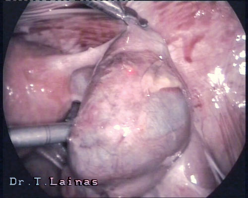
Ectopic pregnancy on right fallopian tube (laparoscopic image).

Depending on the site of implantation, we can distinguish the following types of ectopic pregnancy:

    • Tubal pregnancy (approximately 99% of ectopic pregnancies). The zygote may implant in either of the four segments of the fallopian tube (oviduct): interstitial (3%), isthmic (25%), ampullary (55%) and infundibular (17%). The causes may be, mechanical, functional etc.
    • Ovarian pregnancy: Implantation happens on the ovary and occurs at a frequency 1:7000.
    • Peritoneal pregnancy: Implantation happens in the peritoneum or the intestine either from the beginning or after expulsion of the embryo from the ampulla of the oviduct. It occurs at a frequency 1:8000.
    • Cervical pregnancy: Implantation happens in the inner cervix and occurs at a frequency 1:8000.
    • Simultaneous uterine and ectopic (usually tubal) pregnancy. This the onset of a rare type of multiple pregnancy (1:10000) and occurs more frequently in IVF programs compared to natural conception.

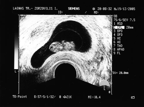
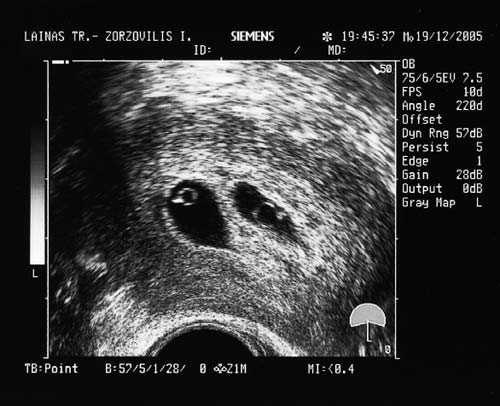
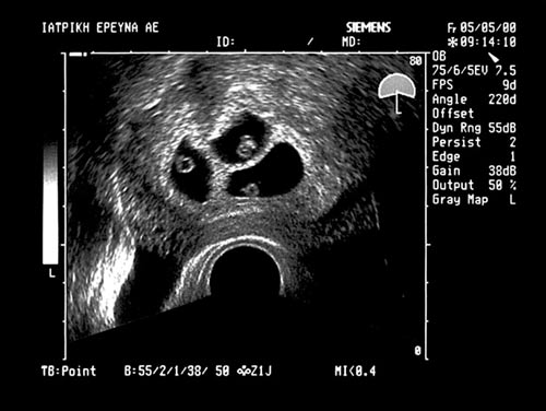
Diagnosis is performed timely and at an early stage (6th week of gestation), as at the first ultrasound examination, that is performed 14 days after a positive hCG test, the exact site of the embryo can be identified.In the past, the diagnosis of ectopic pregnancy was based on symptoms prior to the rpture of the oviduct, and included delay of menstruation, pelvic pain and vaginal bleeding, as well as symptoms after the rupture of the oviduct, which included acute pain and sites of internal bleeding. Nowadays, diagnosis is easy using the levels of beta hCG combined with a vaginal ultrasound.Ectopic tubal pregnancy is treated with laparoscopic surgery, which is now considered the treatment of choice. The traditional laparotomy with removal of the oviduct is a thing of the past, with very few and contradicting indications. Surgical treatment of ectopic tubal pregnancy can be performed by:

      • Retaining of the oviduct (salpingostomy). In this case a longitudinal section of the oviduct is performed using laser CO2, followed by aspiration of the fetus. The oviduct is checked for bleeding, and the wall usually seal by themselves without suturing. It is necessary to measure hCG after a week.
      • Removal of the oviduct (salpingectomy) is performed when there are indications like rupture of large distension of the oviduct wall.

 It is also proposed that the treatment of an ectopic pregnancy can include drug administration, like methotrexate, either intramuscularly or with injection in the embryonic sac under ultrasound or laparoscopic guidance. It is recommended more for cases of interstitial, cervical and peritoneal pregnancy (to induce earlier regression of the placenta, which in these cases is not removed because of the high risk of bleeding).

Management of ectopic pregnancy

Management of ectopic pregnancy using laparoscopic surgery is nowadays the method of choice. Until the 1970s, laparotomy was used due to the inability of early diagnosis, resulting in the rupture of the ectopic pregnancy.

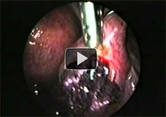
VIDEO: Ectopic pregnancy on right oviduct. Salpingοtomy, fetal removal and retaining of the oviduct.

Early diagnosis is now performed prior to rupture, using beta hCG levels combined with ultrasound examination. Laparoscopic verification is rarely needed. Early diagnosis and laparoscopic surgery have nearly eliminated the disease and mortality due to ectopic pregnancy, and have improved significantly the rate of post-operating infertility.Contraindications for laparoscopic surgery, as reported 20 years ago (Mage G., Canis M., Bruhat M.A) include:

      • Absolute contraindications: interstitial pregnancy, shock, opisthoperitoneal hematocele, and contraindication of general anesthesia.
      • Relative contraindications: hemoperitoneum >15000 ml, obesity, extensive adhesions.


Ectopic pregnancy on both oviducts. Salpingotomy, removal of fetus and retaining of the oviducts.
VIDEO: Ectopic pregnancy on both oviducts. Salpingotomy, removal of fetus and retaining of the oviducts.

Laparoscopic surgical treatment of ectopic tubal pregnancy can be either conservative, with longitudinal salpingotomy, aspiration of the fetus and retaining of the oviduct, or radical, with excision of the oviduct.The choice of laparoscopic treatment in based on criteria, but also on the experience and training of the surgical team. The criteria include history of infertility, prior or repeated ectopic pregnancy, salpingoplasty, site of implantation (isthmus, ampulla, infundibulum), bilateral ectopic tubal pregnancy, and risk of oviduct rupture. Based on the score that results from these criteria it is decided whether to retain or remove the oviduct.a) ) In the case of oviduct rupture, laparoscopic surgery involves a longitudinal section 10-15 mm is above the fetus, removal of the fetus using flushing with saline, aspiration of the fetus away from the wall of the oviduct. The oviduct is examined for bleeding and the wall remains open. Rarely are sutures needed. At the end of the operation the pelvis is flushed with saline, all fetal elements are removed from the pelvis and the other oviduct is examined.Post-operating monitoring includes measurement of hCG levels 2 days after the operation, and then every 2 weeks until hCG is no longer detectable.b) In the case of oviduct removal, the operation is performed using bipolar diathermy for hemostasis, and the excision of the oviduct is done using laser CO2 or scissors. The section is performed very near the oviduct taking care not to damage the blood supply of the ovary.See more: Management of ectopic pregnancy

There are no long term effects for the health of women undergoing IVF. Public worries (usually based on insufficient information), are understandable but are practically have no basis. All the large international epidemiological studies conclude that the risks of developing ovarian, uterine or breast cancer are comparable to the general population.

These large studies from Australia on 30000 women (Lancet 1999), from Great Britain on 5556 women (Human Reproduction 2002), from France on 9255 women (Human Reproduction 2004), as well as other meta-analyses (Nes et al 2002; Kashyap et al 2004) showed that there is not a statistical difference between women who received fertility drugs and developed ovarian, uterine or breast cancer, and women who did not take fertility drugs and developed cancer.

In addition, there is no evidence of increased risk for cancer in children born from IVF, compared to children conceived naturally (study on 17000 vs 30364 children born after IVF; Klip et al 2001).

As a precaution, all women should have a pap-test and a breast examination before the onset of IVF treatment. A mammogram is recommended for women over 35 years old. In cases with breast family history, women should consult a mastologist irrespective of their age.

The rules of regular check up, like gynecological examination, pap-test, ultrasound, breast examination, remain the same, irrespective of whether the woman has undergone or not IVF treatment.

Assisted reproduction cycles are associated with higher rates of multiple pregnancies, the incidence of which has been increased in the last 20 years. In the past, the incidence of a twin pregnancy was 1/80 and of a triplet pregnancy 1/8000, according to the Hellin hypothesis. Today, the increase is approximately 53% for twins and 40% for triplets.

Ultrasound imaging twin pregnancy(EYGONIA file)

The chance of a successful pregnancy after IVF is associated with the number of transferred embryos. At the same time however, there is increased chance that more than one embryos may implant in the uterus, especially for women of young with good quality embryos. Therefore, multiple pregnancy is a side-effect of IVF, and recently there is an international tendency to reduce its occurrence.

Usually, twin pregnancies do not face any problems, provided that there is careful patient monitoring. However, triplet and more high-order pregnancies are more difficult and are associated with more problems and complications, regarding the health of the mother and the chance of premature labour. Premature labour is the most serious reson of perinatal mortality rates and is associated with the number of implanted embryos.


Ultrasound Imaging trigeminal pregnancy (EYGONIA file)

Perinatal death has been reduced recently, due to the technological advancement of neonatal care units and the skilled neonatal clinicians in moitoring and managing premature babies with low birth weight. It is estimated that a mean duration for a twin pregnancy is 260 days, for a triplet pregnancy 210 days and for a quadruplet pregnancy is 190 days, compared to 280 days of a single pregnancy. The development of preeclampsia is 5-10 times higher in multiple pregnancies. The most serious complications of premature labour are brain hemorrhages that lead to paralysis.

For the above reasons, it is vital to restrict the number of transferred embryos.

The is a rare chance of natural twinning after IVF, after splitting of a single embryo into two separate embryos, giving rise to mono-oogenic twins.

Our team has designed and applies a novel approach for the management of established severe OHSS. Using this method we safely and effectively achieved the regression of the syndrome, on an outpatient basis, and completely avoided the need for hospitalization in all patients. Using this method, all high risk women can safely proceed at least to oocyte retrieval, avoiding a cycle cancellation.

Until today, the only safe way to prevent OHSS was withholding hCG and cycle cancellation in high risk women, which were associated with physical, psychological and financial burden. Our novel method challenges this dogma of cycle cancellation. Our study has been described as novel and pioneering by the international scientific community.

In addition, using this method we achieved the first live births of healthy babies worldwide following blastocyst transfer in women with established severe OHSS.

Our new approach is described in 3 publications in the journal Reproductive Biomedicine Online.

Live births after management of severe OHSS by GnRH antagonist administration in the luteal phase. TG Lainas, IA Sfontouris, IZ Zorzovilis, GK Petsas, GT Lainas, E Alexopoulou, EM Kolibianakis
RBMOnline - Vol 19 No 6. 2009 789-795
See the publication

Management of severe OHSS using GnRH antagonist and blastocyst cryopreservation in PCOS patients treated with long protocol. TG Lainas, IA Sfontouris, IZ Zorzovilis, GK Petsas, GT Lainas, GS Iliadis, EM Kolibianakis.
RBMOnline - Vol 18 No 1. 2009 15-20
See the publication

Managment of severe early ovarian hyperstimulation syndrome by re-initiation of GnRH antagonist. TG Lainas, IA Sfontouris, IZ Zorzovilis, GK Petsas, GT Lainas, EM
Kolibianakis
RBMOnline - Vol 15. No 4. 2007 408-412
See the publication

We have published the largest study in the literature for women with PCOS undergoing IVF. Our results show that the GnRH antagonist protocol is associated with a significantly lower incidence of OHSS compared to the long protocol, while maintaining high pregnancy rates. Therefore, the antagonist protocol can be the protocol of choice for PCOS women undergoing IVF.

Flexible GnRH antagonist protocol versus GnRH agonist long protocol in patients with polycystic ovary syndrome treated for IVF: a prospective RCT. Trifon G Lainas, Ioannis A Sfontouris, Ioannis Z Zorzovilis, George K Petsas, George T Lainas, Efthymia Alexopoulou, Efstratios M Kolibianakis Human Reproduction, 2010, Vol 25, No 3, pp. 683-689 See the publication

In another study of our team, we used a different antagonist protocol (D1), which was also associated with a lower chance of OHDD development. The study included women with PCOS and compared the long protocol with an antagonist protocol in which antagonist was initiated on day 1 of ovarian stimulation, instead of day 6.

Initiation of GnRH antagonist on Day 1 of stimulation as compared to the long agonist protocol in PCOS patients. A randomized controlled trial: effect on hormonal levels and follicular development. Lainas TG, Petsas GK, Zorzovilis IZ, Iliadis GS, Lainas GT, Cazlaris HE, Kolibianakis EM Human Reproduction, 2007, Vol. 22, No 6, pp. 1540-1546 See the publication

We also use methylprednisolone for the prevention of OHSS in high risk patients. Using this method we successfully treated all cases of OHSS at an outpatient level. Patients with severe OHSS who were hospitalized were very few.

Administration of Methylprednizolone to prevent severe ovarian hyperstimulation syndrome in patients undergoing in vitro fertilization. T. Lainas et al., Fertil. Steril. 2002;78(3):529-533). See the publication

OHSS is a serious complication of ovarian stimulation in IVF. It can be mild, moderate or severe, depending on the severity of symptoms. Symptoms include abdominal distension, stomach ache, vomiting, increase of body weight and reduced urination. Moderate OHSS occurs in 3-6%, while severe OHSS occurs in 0.2-1% of women undergoing IVF. Mild OHSS lacks clinical significance, while severe OHSS includes fluid accumulation in the third space, increase of ovarian volume, hematocrit, white blood cells and liver indices. More rarely in critical forms there might be breathing difficulty, fainting and abnormal hematological and biochemical markers. In this case hospitalization is necessary.

Risk factors for the development of OHSS include young age, low body weight, high gonadotrophin doses, previous OHSS history, high dose of hCG and polycystic ovaries.

Women with polycystic ovaries are at greater risk for developing OHSS (9-38%). This variation is mainly due to the lack of a universal classification system of OHSS.

OHSS is caused by the exogenous hCG administration in the presence of a large number of follicles (more than 20).

OHSS develops 3-7 days after oocyte retrieval (early onset), or can be pregnancy-induced, 12-17 days after oocyte retrieval (late onset).

The pathophysiological cascade of events is as follows: Ovarian stimulation using exogenous gonadotrophins is followed by multiple follicle development. Administration of hCG to trigger final oocyte maturation causes massive luteinization. Secretion of angiogenic factors, such as VEGF, by the multiple corpora lutea leads to increased permeability of blood vessels and shift of fluid to the third space. The clinical evidence of OHSS is the presence of ascites.

A subsequent pregnancy is not threatened by OHSS. But pregnancy can further induce the symptoms of OHSS.

OHSS can be prevented and treated

Accurate prognosis, active prevention and drastic treatment form an effective approach for the management of OHSS. The scientific team of Eugonia has a long experience and accumulated knowledge on risk factors and prevention of OHSS, as shown by our everyday clinical practice, and also our published studies in international journals. Our earlier studies are related to OHSS prevention (using methylprednisolone) or the use of GnRH antagonist protocols. Our latest studies report a novel treatment of established severe OHSS, which is a safe and effective solution that can change the philosophy of OHSS management.

Eugonia - Assisted Reproduction Unit
Konstantinou Ventiri 7(HILTON), 11528 Athens

  • Email: info@eugonia.com.gr
  • Τel.: +30 210 723 6333
  • Fax: +30 210 721 3623

Member of:

Google Reviews

Photo gallery

© Copyright 2025 Eugonia - Assisted Reproduction Unit in Greece Developed by NetValue. All rights reserved.
Menu