Menu

Blog

These protocols refer to the pharmaceutical regulation in order to achieve controlled ovarian stimulation (COS). The reason for their use is multiple follicle development and avoidance of early follicle rupture due to the surge in LH. Early follicle rupture is equivalent to the loss of oocytes prior to their scheduled pick-up (oocyte retrieval) that is performed by the gynaecologist under ultrasound guidance.

The decision of which stimulation protocol to use depends on the individual nature of each patient's cycle, the ovarian response in previous cycles, the age and other factors assessed by the doctor.

Nonetheless, the application of these protocols is flexible as every patient may respond to the drugs differently. An important part of our task is to define the best therapeutic protocol and decide on the ideal dosage for each separate case.

The most common protocols are:

This category includes women that show an excessive response to ovarian stimulation drugs which results to an excessive recruitment and multiple follicle development.

The factors that favour excessive ovarian response are polycystic ovaries, young age, reduced body weight (low BMI; body mass index), high gonadotropin doses or progressively increasing dose, aggressive stimulation protocols, excessive ovarian response in a previous IVF treatment cycle, etc.

Hyperesponders have increased chances of developing OHSS (ovarian hyperstimulation syndrome) and possibly the severe form of the syndrome, which is one of the most serious complications of IVF treatment.

The scientific team of Eugonia has shown international innovation and experience in the prevention and management of OHSS with a significant number of studies published in internationally acclaimed scientific journals and lectures of the Head of the Eugonia Unit, Dr T. Lainas.

The publications reflect the knowledge and experience applied in everyday clinical practice.

Prevention of OHSS

The publications related to OHSS prevention are either on corticoids administration (also an older opinion of our Unit), or on the use of antagonist protocols which are thought to be safer for hyperesponders.

Also see: Antagonist protocol.

See our publications:

Live births after management of severe OHSS by GnRH antagonist administration in the luteal phase.

Management of severe OHSS using GnRH antagonist and blastocyst cryopreservation in PCOS patients treated with long protocol.

Management of severe early ovarian hyperstimulation syndrome by re-initiation of GnRH antagonist.

Management of OHSS

The proposal of the scientific team of Eugonia for antagonist administration in the luteal phase for the management of already established severe OHSS is a world's first.

See: our news

In the IVF treatment programs, the term "poor responder" is commonly mentioned. This means that the ovaries of these women show significantly reduced response to the ovarian stimulation drugs that are administered for the recruitment and development of multiple follicles. When compared with the population of women that show normal ovarian response, poor responders produce fewer eggs and have lower pregnancy rates in IVF treatment cycles.

Unfortunately, there is no internationally accepted definition of poor response. The majority of doctors agree that this cohort, includes women that during an IVF cycle recruited and developed less than 3 or 5 follicles and produced less than 3-5 mature eggs even when high gonadotrophin doses and various ovarian stimulation protocols were used.

Various researchers have described numerous criteria, such as: low E2 levels during stimulation, increased FSH levels, low levels of AMH on the 3rd day of the menstrual cycle, at least one cancelled cycle, no response to increased gonadotropin doses, long stimulation duration etc.

High gonadotropin doses (>300IU) exclude the chance of a reduced dosage that may be linked to the small number of follicles that will be recruited and develop.

Another result of the small number of follicles that develop in the ovaries is the low E2 levels during stimulation.

Poor response is due to reduced ovarian reserve. The ovaries are unable to produce more eggs than the ones they already have, even if they are bombarded with high gonadotropin doses.

It is self explanatory that from a small number of follicles, we will retrieve a small number of eggs. If for example the number of eggs collected is 5, only a percentage of these will be mature (i.e. able to fertilise). Only a percentage of these mature eggs will fertilise, divide and have a good implantation potential.

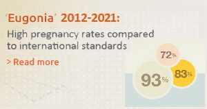
Such loses significantly lower the number of embryos available for transfer. For this reason, poor responders show lower pregnancy rates when compared to women with a normal response.

The factors responsible for poor response may be genetic and environmental, as well as ovarian surgery, endometriosis, immunological, auto-antibodies, reduced number of FSH receptors on granulosa cells, unknown, etc. Ofcourse, the main factor responsible is a reduced ovarian reserve.

Poor responders are not necessarily subfertile when there is no other cause of subfertility.

Prediction of poor response is possible with: a) a basal antral follicle count (e.g. <5 basal antral follicles) by transvaginal ultrasound, b) increased FSH levels (e.g. >12) on the 3rd day of the cycle, c) low AMH levels (e.g. <0.3-0.6 ng/ml). In the past, inhibin-B and GnSAF were used. In IVF treatment, it is especially useful to record the ovarian response to high gonadotropin doses (>300IU) in any previous cycles.

Management of poor responders

Unfortunately, international scientific literature (Tarlatzis, Ubaldi) shows that there is no ideal stimulation protocol.

There have been described and compared: high gonadotropin doses (>300IU), the use of recombinant gonadotropins versus urinary gonadotrophins, administration of gonadotropins in the luteal phase, short and ultra short GnRH agonist protocols with a reduction in the GnRH agonist dose (Arvekap, Daronda, Suprefact), long protocol with agonist administration in the luteal phase, GnRH antagonist protocol, growth hormone (GH) administration, gluco-corticoids administration, use of contraceptives, aromatase inhibitors, DHEA (dehydroepiandrosterone), use of only intracytoplasmic sperm injection (ICSI) for fertilization, assisted hatching, natural cycles, modified natural cycles (M.N.C.) and finally oocyte or embryo donation

Our contribution for poor responders

We have published the largest study in the international literature for poor responders that underwent IVF treatment. Our study was published in the internationally acclaimed scientific journal of Human Reproduction, which is the official journal of the European Society for Human Reproduction and Embryology (ESHRE). It is a prospective randomized study that shows that the flexible GnRH antagonist protocol correlates with statistically higher ongoing pregnancy rates when compared to the short GnRH agonist protocol. In conclusion, the antagonist protocol may be considered as the protocol of choice for women with poor ovarian response that undergo IVF treatment.

See our publication

AMH is a dimeric glycoprotein and a member of the transforming growth factor β (TGF-β) family. In men, it is produced by the Sertoli cells of the testes and its main action is in the regression of the Müllerian duct in male fetuses during sexual differentiation (8th-10th week of their embryonic life).

In women, AMH is produced by small follicles (<6 mm) in the ovaries. AMH production is gradually reduced as the follicle grows in diameter and it is practically non-traceable in follicles >8 mm.

A large number of studies suggest that AMH greatly reflects the total number of follicles that remain in the ovaries, i.e. it is a powerful indicator of ovarian reserve and a prognostic factor of ovarian response in an assisted reproduction treatment cycle to follow. Researchers also support that AMH measurement is equally reliable to the ultrasound assessment of the number of small antral follicles in the ovaries, which is the principal prognostic factor of ovarian reserve. AMH levels are reduced as the age of the woman increases and as the ovarian reserve decreases, while at the same time there is a simultaneous reduction in the number of developing follicles visible on ultrasound. On the contrary, in cases of a large number of follicles, as with women with polycystic ovaries, AMH levels are excessively increased.

It has also been suggested that the AMH blood levels are a predictive factor for the chances of success in an assisted reproduction treatment cycle to follow, although opinions on this do vary.

The normal values of AMH may vary and they depend of the method and the laboratory that the measurement took place. According to scientific literature and our experience, AMH values are classified as follows:

AMH Value Interpretetion
> 3.0 ng/ml High levels (usually in polycystic ovaries)
> 1.0 ng/ml Normal
0.7 - 0.9 ng/ml Lower normal range
0.3 - 0.6 ng/ml Low levels
< 0.3 ng/ml Very low levels

At Eugonia, in line with new scientific developments, we use the AMH measurement in combination with ultrasound assessment of the ovaries (number of small antral follicles and ovarian volume) and FSH and E2 levels, in order to determine ovarian reserve and select the optimal stimulation protocol.

Oestradiol (Ε2 or 17-β oestradiol)

Oestradiol is a steroid hormone, mainly produced in the gonads (ovaries and testes). It is one of the major oestrogens in women of reproductive age.Men also produce oestradiol as a by-product of testosterone metabolism. Oestradiol levels in men generally fall in the same range as those of postmenopausal women.Apart from its functioning as a sex hormone, oestradiol has a beneficial effect in other tissues such as bones.Oestradiol, in women of reproductive age, is produced by granulosa cells within the follicles of female ovaries.In males oestradiol is produced in lower levels, in the adrenal glands and testes.

Oestradiol Measurement

Serum oestradiol measurement in women reflects primarily the activity of the ovaries. Serum oestradiol measurement during infertility treatment reflects follicular growth. Elevated levels of oestradiol  and other oestrogens are correlated with oestrogen producing tumors. Elevated oestradiol levels also occur in precocious puberty.Oestradiol and FSH blood tests  are performed on the 2nd or 3rd day of the menstrual cycle in order to test the ovarian reserve.  A normal FSH value (<10) may be mistakenly considered normal if oestradiol levels are high (>60pg/ml).

FSH (Follicle stimulation hormone) is a hormone that stimulates the ovary to produce oocytes. FSH is produced from the pituitary (or hypophysis). The pituitary is an endocrine gland in the base of the brain that secretes hormones for the control of the function of the thyroid gland, the adrenal glands, the gonads (testes-ovaries) and it also secretes growth hormone and prolactin. For the function of the gonads, the pituitary secretes two hormones that are called gonadotropins. The follicle stimulating hormone (FSH) promotes the development of the follicles, and the luteinising hormone (LH) promotes steroidogenesis in the ovary and is responsible for the final maturation of the oocyte, the ovulation and the beginning and continuation of the function of the corpus luteum.

The two gonadotropins FSH and LH are produced and stored in the gonadotroph cells of the pituitary and they exert their biological action on the gonads (testes-ovareis) via their receptors located on the target organs.

The FSH and LH secretion is controlled by the GnRH hormone, which is secreted in pulses by the pituitary. Thus, the FSH and LH gonadotropin secretion also takes place in pulses. As we have seen, the pituitary is under control of the hypothalamus, through the GnRH. On the contrary, the hormone secretion from the pituitary and the hypothalamus is affected by the levels of ovarian hormones. Therefore, there is a hypothalamic-pituitary-ovarian hormone axis, with a mechanism of negative feedback. Oestradiol (E2) produces in the ovary has a negative feedback on the on the FSH secretion and a positive feedback on the LH secretion.

Measurements have shown that the GnRH levels are lower in children and higher in adults. GnRH levels are also increased during the middle of the menstrual cycle (ovulation). GnRH levels are higher in a post-menopausal woman that in a woman during her reproductive age. Similar to that is the course of the gonadotropin levels. The post-menopausal period in a woman is characterized by almost the cancellation of the ovarian function and an excessive rise in the FSH and LH secretion by the pituitary.

The rise of FSH and LH levels during menopause is not sudden, as when the ovaries are removed. A period of gradual reduction of the ovarian function, i.e. a period of reduced fertility whose last stop is menopause, has preceded. During this time increased FSH levels are observed.

It thus appears as an effort of the pituitary through the continuously increasing gonadotropin levels to stimulate the ovary by recruiting and developing follicles in order to preserve fertility. The ovary however has a reduced reserve and cannot effectively respond to the pituitary commands.

In conclusion, increased FSH levels show reduced ovarian reserve and increased biological ovarian age.

This is about counting the number of follicles with an antrum (antral follicles) during the basic ultrasound. These follicles, also known as small antral follicles, are of small size (2-8mm) and can be counted in the basic ultrasound on the 2nd or 3rd day of the menstrual cycle.

Recent studies and long clinical experience have shown that the counting of the antral follicles is a strong prognostic factor of estimating ovarian reserves, since it greatly reflects the reserves of the primordial follicles (i.e. the total of the very small follicles, not visible during the ultrasound) of the ovaries. With the application of a complicated statistical analysis (multivariate logistic regression analysis) researchers have shown that the number of small antral follicles in the basic ultrasound, in combination with the age of a woman, is the best predictive factor for the follicles that are going to develop after pharmaceutical stimulation of the ovaries.

Every primordial follicle contains an immature oocyte, which has the potential to develop and mature at some point in the future, during the reproductive life of a woman. Therefore, when there is a small number antral follicles in the basic ultrasound, this means that there is an equally low number of primordial follicles (i.e. low ovarian reserve). On the contrary, an excessive number of antral follicles is an indication for polycystic ovaries.

The assessment of the ovarian reserve with a transvaginal ultrasound counting of the small antral follicles in combination with the measuring of the FSH, E2 and AMH offers important information for a future assisted reproduction treatment cycle, such as:


  • The ovarian response to the pharmaceutical stimulation, i.e. the number of follicles that are going to develop. The prediction of the ovarian response is more precise when there are very low or very high numbers of small antral follicles. On the contrary, an average number of small antral follicles is a less powerful prediction factor.
  • The number of oocytes that are going to be retrieved.
  • The chances of cycle cancellation.
  • The optimal stimulation protocol.
  • The quality of the oocytes and the embryos.
  • The chances of achieving a pregnancy.

The term ovarian reserve refers to the reserves of the ovaries in follicle numbers, which is expressed with the ability of the ovaries to recruit follicles when they are stimulated with drugs. As the age advances, the ovarian response to exogenous gonadotropins (e.g. Puregon, Gonal, Altermon, Menopur etc.) is reduced.

The aim of estimating the ovarian reserve is to calculate in a reliable way the number of follicles and therefore the number or oocytes that remain at a given time in the ovaries of a woman.

The precise evaluation of the ovarian reserve is especially useful in assisted reproduction treatment programs because it aids the prediction of the ovarian response to exogenous gonadotropins. Therefore, one can calculate reliably the number of follicles that are going to develop from the ovary following the administration of the optimal pharmaceutical protocol and the appropriate dosage.

The number of oocytes that will be retrieved depends on the number of follicles that are going to develop and it correlates with the chances of achieving a pregnancy. Thus, the right assessment of the ovarian reserve offers a reliable and precise prediction of the pregnancy success rates.

Assessment methods

The usual tests for the evaluation of the ovarian reserve is the hormone testing for FSH, E2 and the transvaginal ultrasound for the measuring of the ovarian volume and mainly counting the number of the small antral follicles on the 2nd or 3rd day of the menstrual cycle.

Follicles and age

The oocyte production of a woman begins during the embryonic development of the ovary. At this stage, the primordial germ cells that are called oogonia multiply fast with mitotic divisions. In the 20th week of embryonic development, the oogonia reach their maximum number of 7 million per ovary. From this time onwards, their number decreases dramatically through the process of atresia. Thus, at birth germ cells are estimated around 1-2 million, whilst at puberty around 300-400,000 remain. This reserve does not renew itself, but it gradually reduces as the age of the woman advances. At the age of 37 years, there are around 25,000 primitive follicles, while at menopause these reduce to about 1000, which disappear by 71 years of age.

It is possible that a small percentage of women undergoing ovarian stimulation for fertility treatments may have to be cancelled. Cancellation of a treatment cycle is recommended when the response of the ovaries to the drugs is contrary to our expectations. Our aim is to always give patients the best chances of success in each cycle undertaken and to eliminate the already minimal risk of possible complications.

Cancellation during treatment cycle is usually recommended in the following cases:

During the phase of stimulation with the use of gonadotropins, it is possible to see reduced ovarian response. If following the correction of the daily gonadotropin dose by the doctors of the Unit, the ovarian response is still low or non-existent, cancellation of the cycle and start of a new treatment cycle with a different protocol or a higher starting dose of gonadotropins is recommended.

If the ovarian response is considered to be higher than certain limits during monitoring, there is a real danger of developing severe ovarian hyperstimulation syndrome that can pose a great risk for patient's health. This is more common in women with polycystic ovarian syndrome, who show a certain response according to the gonadotropin dose. If the dose is below a certain level there is no response and if the dose is above that level this results in hyperstimulation. It is therefore preferable to cancel a cycle, despite the increased negative phychological effect that this may entail for the patient, instead of presenting the patient with an increased risk of developing severe ovarian hyperstimulation syndrome.

A new treatment cycle can then be undertaken in a different menstrual cycle, with a different dosage and probably a change in the protocol used.

During the long protocol, it is possible to cancel the cycle during the suppression phase, either due to the appearance of a functional ovarian cyst which reforms following aspiration, or due to the inefficient suppression of ovarian function.

In the case of protocols for the transfer of frozen thawed embryos, the cancellation of the cycle is recommended when the thickness or the echo pattern of the endometrium is deemed unsatisfactory.

Also, it is possible for the treatment cycle not to reach its final stage (embryo transfer) for various reasons such as failed egg collection (when no oocytes are collected), fertilization failure, no embryo cleavage, or even zero survival of frozen embryos following their thaw. However, the chances of any of that happening are quite low (about 1-4% in all cycles, depending of the age of the woman).

The day when sufficient follicle maturation is observed you will be informed of the exact time for your last injection which will induce final oocyte maturation and ovulation (Pregnyl or Profasi or Ovitrelle) as well as the dose that must be used. This injection is human chorionic gonadotrophin which mimics the LH action and induces final oocyte maturation and ovulation. The injection is administered late in the evening or after midnight. Ovulation is expected about 36 hours later.

This last injection concludes the painful phase of daily injections and marks the end of GnRH analogue and gonadotrophin administration. The following day is a day to rest and prepare for the egg collection. This last injection is very particular in that it must be done at a very specific time, which will be the deciding factor for the time of egg collection. If we attempt to retrieve the eggs before they develop completely, i.e. much earlier than 36 hours after the injection, we shall collect immature eggs. On the contrary, if the injection is done before the programmed time there is a danger that ovulation will occur prior to the egg collection and the eggs will be lost.

If the injection is not done, the egg collection will have to be cancelled or postponed. The eggs will not be mature and it will not be possible to recover them from the ovary. If the injection is not done you must inform our Unit the following morning so your egg collection is re-programmed and the entire cycle is not cancelled.

Eugonia - Assisted Reproduction Unit
Konstantinou Ventiri 7(HILTON), 11528 Athens

  • Email: info@eugonia.com.gr
  • Τel.: +30 210 723 6333
  • Fax: +30 210 721 3623

Member of:

Google Reviews

Photo gallery

© Copyright 2025 Eugonia - Assisted Reproduction Unit in Greece Developed by NetValue. All rights reserved.
Menu