Menu

Blog

Mature oocyte at the stage of metaphase II.
Video: Intracytoplasmic sperm injection (ICSI)

After the retrieving the sperm sample from the male partner, the sample is prepared to isolate the normal motile spermatozoa. Even a few thousand spermatozoa are enough for ICSI to be performed. Hoverer, in cases with immotile sperm of obstructive azoospermia, the sperm must be retrieved using surgical methods from the epididymis or the testis.

Micropippetes used for ICSI

ICSI is performed in two steps. First, the oocytes are stripped from surrounding cumulus cells using a combination of enzymatic (hyaluronidase) and mechanical methods. This step is important for the identification of mature oocytes and for the esae of micromanipulations. Maturity is determined by the presence of the first polar body. Only mature oocytes can be used for ICSI. Immature ones cannot be used.

Procedure of ICSI
Procedure of ICSI.

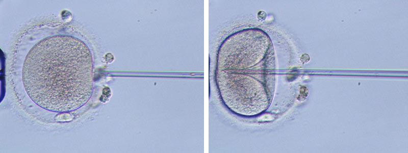
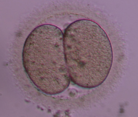
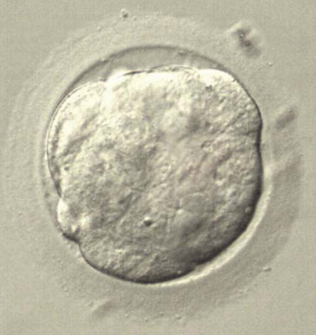
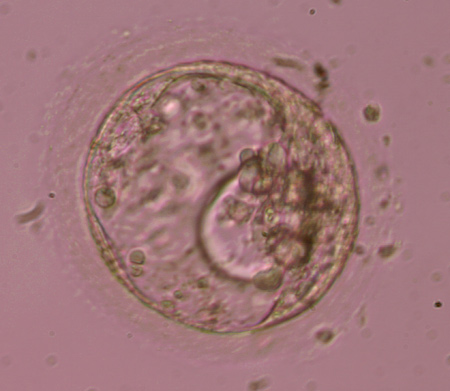
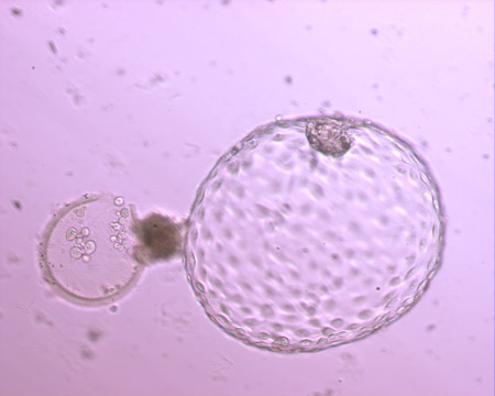
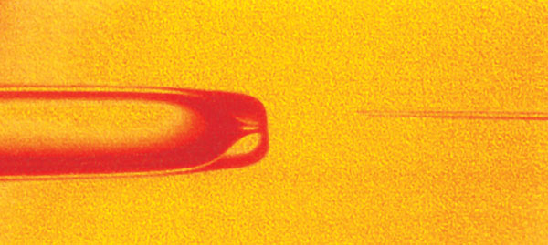
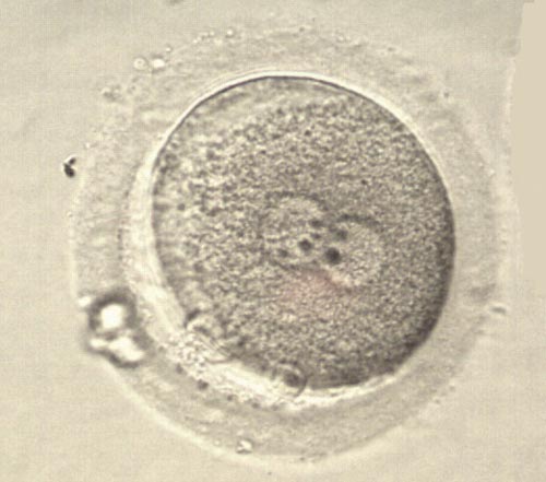
A. Mature oocyte with extruded first polar body (at 6 o'clock). The injection pipette that contains the spermatozoon approaches the oocyte. (Eugonia archive)

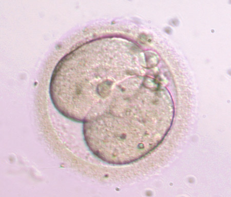
B. The injection pipette has penetrated the zona pelucida and the oocyte membrane. The spermatozoon will be deposited inside the oocyte cytoplasm. (Eugonia archive)


The second step is the actual performance of ICSI. The naked oocytes are placed in drops of culture medium in a petri dish and is help in place by the holding pippete. In a different drop, sperm are deposited, and each spermatozoon is immobilized and aspirated in to the special injection pipette. The spermatozoon is then injected inside the oocyte (one spermatozzon per oocyte). Each injection usually lasts for a few seconds, but the entire procedure may take several hours to complete in cases of high oocyte numbers or extremely low sperm numbers.

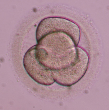
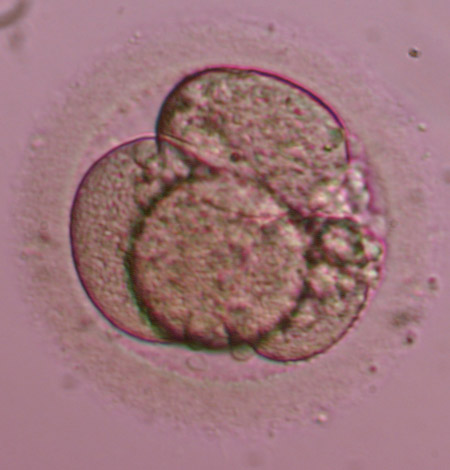
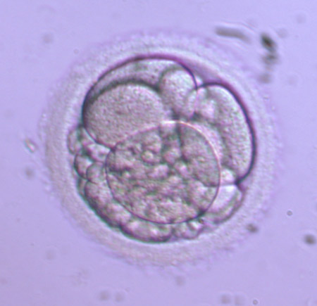
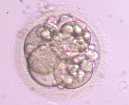
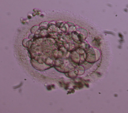
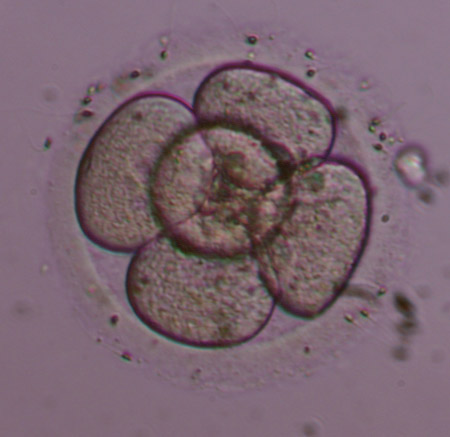
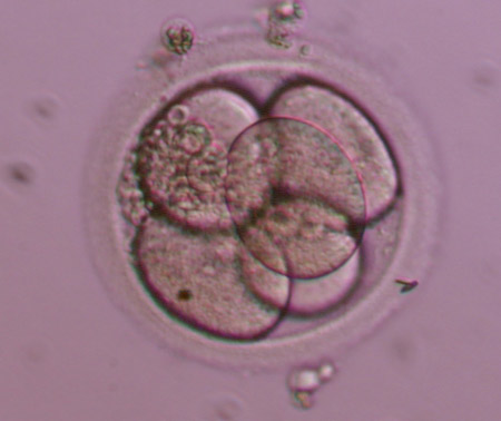
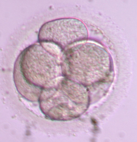
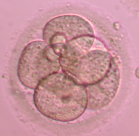
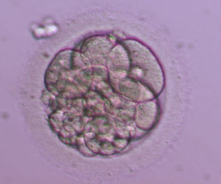
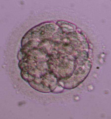
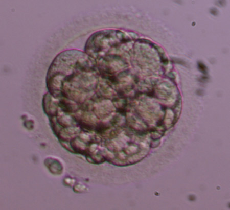
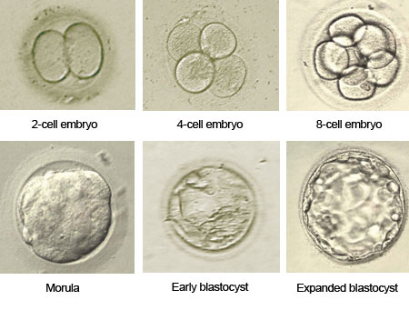
After the injection, the oocytes are placed back in the incubator. Polyspermy is avoided using this method. Fertilization rates are usually high after ICSI (higher than 60%). Fertilization failure after ICSI is usually related to inability of the sperm chromatin to decondense, or abnormal oocyte activation. Damage rates are usually low (2-3% in experienced hands). From that point on, the procedure is the common irrespective of the fertilization method (i.e. formation of pronuclei, cleavage into 2,4,8, etc blastomeres, and embryo transfer).

IVF, apart from infertility treatment, has also a diagnostic value as it gives an indication that the oocytes and sperm of a couple can interact. In some cases of unexplained infertility we discover a possible cause for infertility and may obtain useful information, such as no attachment of sperm on the zona pelucida, low sperm motility after a few hours of culture etc. The laboratory conditions and culture media are optimized so that they do not affect fertilization in any way.

Fertilization rates do not reflect the quality of resulting embryos and a low fertilization rate should not cause worries about the outcome of IVF.

Usually, normal fertilization rates in the laboratory range from 50% to 100%. It depends on:

  • The quality and maturity of the oocytes
  • The fertilizing ability of the sperm
  • Optimal culture conditions

Polyspermy rates are low (3-5% of oocytes).

A proportion of the inseminated oocytes after IVF or ICSI either do not form pronuclei, or form only I pronucleus (1PN) or multiple pronuclei (3PN, 4PN). After conventional IVF, the presence of multiple PNs means that the oocyte was fertilized by more than just one spermatozoa. These embryos are called polyspermic and have surplus numbers of chromosomes. Polyspermic embryos should not be transferred as they are related in abnormal pregnancies (miscarriages, myli pregnancy).

Oocyte with normal fertilization with 2 pronuclei and 2 polar bodies.

The following morning after oocyte retrieval, i.e. 16-20 hours after insemination the fertilization check is performed. The embryologists check under the microscope how many oocytes have been fertilized normally and isolate unfertilized or abnormally fertilized oocytes. Normal fertilization is verified by the presence of 2 pronuclei (2PN) and 2 polar bodies.

Schematic representation of the fertilization process.

The chromosomes of the spermatozoon and the oocyte have been condensed and are enveloped by a membrane, giving rise to the two pronuclei (1 male and 1 female). At the same time the second polar body is extruded marking the completion of meiosis II. The first cells of the new embryo is now called zygote and has the same size and shape as the oocyte.

During natural conception, the blastocyst hatches from the embryonic shell, the zona pellucida, on the 5th or 6th day after fertilization. The fully hatched blastocyst is the last free-form embryonic stage and the only stage when the embryo has the capacity to attach and implant in the endometrium. However, in some cases, the zona pellucida is harder or thicker than normal, obstructing the process of hatching and, as a result, impairing successful implantation.

Hatching blastocysts after thawing.
Following an artificial opening on the zone pelloucida using laser the blastocysts have started to hatch.

When the embryos develop in culture, there is another possible intervention before the embryo transfer; the embryologist can assist blastocyst hatching by opening a small hole on the zona pellucida (assisted hatching), using either a special laser device or a chemical solution.

The initial excitement over the usefulness of assisted hatching in implantation has not been widely accepted by embryologists. The method does not seem to significantly increase implantation rates, while it subjects the embryos to further stress. However, assisted hatching has been proven to slightly improve implantation in special cases, such as thick zona, frozen-thawed embryos, eggs from women of increased age, etc.

Eugonia offers the assisted hatching method with the use of laser, in the cases that is has been deemed necessary by our scientific team.

Embryo development begins a few hours after the formation of the zygote. The zygote divides in two cells that are called blastomeres. The next divisions follow, giving rise to more and smaller blastomeres. This process is called cleavage.

Stages of embryo development until the blastocyst stage

Two days after oocyte retrieval cleavage has started and embryos have undergone 2 cell divisions. Cleavage rates are high, about 95%. Typically, the first division occurs about 16 hours after fertilization (2-cell embryo). The second division happens 12 hours later (4-cell embryo) and divisions continue in an increasing rate. Between 4 and 8-cell stage the embryonic genome is activated. The next stages are the morula and the blastocyst.

Correct cleavage is important for embryo quality. The number and morphology of blastomeres, the presence of fragmentation, the morphology and fusion of pronuclei and the synchronous divisions are important features for the evaluation of embryos and are related to IVF outcome.

Therefore, embryos with late cleavage (e.g. 2-cell stage on day 2) are considered inferior to 4-cell stage embryos.

The development of the zygote up the blastocyst stage

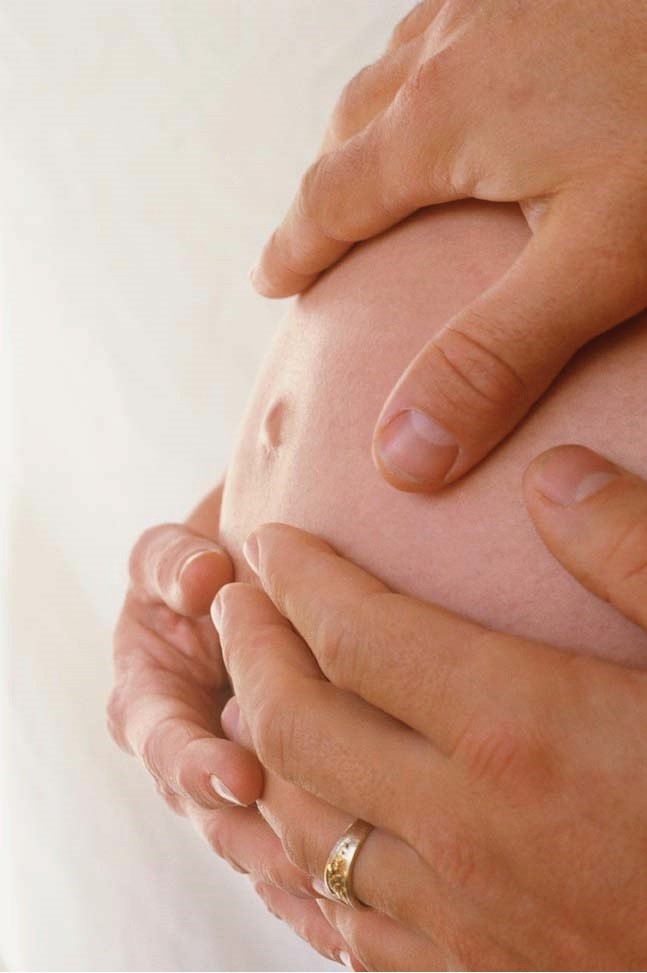
Surrogacy applies to couples with normal eggs and sperm but the woman has a non functional uterus or is unfit to gestate for medical reasons.

Fertilization occurs in the embryology laboratory.

The gestation of a couple's embryo by a third woman is allowed by judicial permission according to article 1458 of N.3089/02, provided that there is a documented and unselfish agreement between the couple and the surrogate mother (and her husband if the latter is married).


Day 2 embryos

2 cell Grade I
2 cell Grade I
2-cell Grade II
2-cell Grade II
2-cell Grade III
2-cell Grade III
 
3-cell Grade II
3-cell Grade II
 
4-cell Grade I
4-cell Grade I
4-cell Grade II
4-cell Grade II
4-cell Grade II-III
4-cell Grade II-III
4-cell Grade III
4-cell Grade III
4-cell Grade IV
4-cell Grade IV
 

Day 3 embryos

5-cell Grade I
5-cell Grade I
5-cell Grade II
5-cell Grade II
6-cell Grade I
6-cell Grade I
6-cell Grade I-II
6-cell Grade I-II
6-cell Grade II-III
6-cell Grade II-III
6-cell Grade III
6-cell Grade III
6-cell Grade IV
6-cell Grade IV
 
7-cell Grade II
7-cell Grade II
7-cell Grade II-III
7-cell Grade II-III
8-cell Grade I (fertilized with ICSI)
8-cell Grade I  (fertilized with IVF)
8-cell Grade I (fertilized with IVF)
8-cell Grade II
8-cell Grade II
8-cell Grade II-III
8-cell Grade II-III
8-cell Grade III
8-cell Grade III
8-cell Grade IV
8-cell Grade IV
9-cell Grade I
9-cell Grade I
 
12-cell Grade I
12-cell Grade I
 

Day 4 embryos

Morula
Morula
 

Day 5/6 embryos (Blastocysts)

Early blastocyst
Early blastocyst
 
Full blastocyst 3ΑΑ
Full blastocyst 3ΑΑ
Full blastocyst 3ΑΒ
Full blastocyst 3ΑΒ
Full blastocyst ΒΒ
Full blastocyst ΒΒ
Full blastocyst 3CC
Full blastocyst 3CC
Expanded blastocyst 4AA
Expanded blastocyst 4AΒ
Expanded blastocyst 4AC
Expanded blastocyst 4BB
Expanded blastocyst 4BC
Expanded blastocyst 4CC
Hatching blastocyst 5AA
Fully hatched blastocyst 6ΑΑ

The achievement of a successful pregnancy largely depends on the number and quality of embryos transferred to the uterus. Therefore, we need to evaluate a patient's embryos during their development and select for transfer those with the highest morphological characteristics.

Evaluation of Day 2-3 embryos

Cleavage

On Day 2 after oocyte retrieval the embryos must have 2-4 cells, with optimal stage the 4-cell stage. On Day 3, the embryos must have 5-8 cells, with 8-cell embryos being the best developmentally.

Morphology- Fragmentation

Embryos are categorized into four grades (I-IV) based on size and shape of blastomeres and percentage of fragmentation. Grade I embryos are the best morphologically, without fragmentation, normal size and shape of blastomeres. On the contrary, Grade IV includes poor quality embryos with total fragmentation or degenerated blastomeres. Intermediate Grades are II and III. Ideally, on day 2 we want to have 4-cell embryos with grades I or II. ON day 3, best embryos are regarded 8-cell embryos with grades I or II.

4-cell embryos

4-cell embryos
 

8-cell embryos

8-cell embryos
8-cell embryos

Other criteria

Multinucleation

Normally, each blastocyst of a developing embryo must have one visible nucleus. The presence of multiple nuclei (multinucleation) indicates an abnormal number of chromosomes and is related with a lower chance of implantation.

Uniformity of blastomeres

Uniformity of blastomeres is also related to embryo quality. Normally, a good quality embryo has blastomeres of equal size and shape. However, it is normal for a blastomere to have a larger size than the rest for odd-number cell stage (3, 5,7 cells).

Cytoplasm

The presence of granules or vacuoles inside the blastomeres should be recorded, although these characteristics have not been definitely related to poor embryo quality.

Thickness of zona pelucida

The zona pelucida is an acellular glycoprotein layer that surrounds preimplantation embryos. During expansion of the blastocyst, the zona ruptures and allows the embryo to hatch and implant. Increased thickness of the zona pelucida is related to lower chances of implantation as they embryo may get trapped inside it and fail to hatch successfully.

Evaluation of blastocysts

See: Blastocysts.

Metabolic criteria

See: Metabolomics.

Eugonia - Assisted Reproduction Unit
Konstantinou Ventiri 7(HILTON), 11528 Athens

  • Email: info@eugonia.com.gr
  • Τel.: +30 210 723 6333
  • Fax: +30 210 721 3623

Member of:

Google Reviews

Photo gallery

© Copyright 2025 Eugonia - Assisted Reproduction Unit in Greece Developed by NetValue. All rights reserved.
Menu