

During the last few years, ovarian cysts are treated with laparoscopic surgery with excellent post-operative results. Thorough assessment precedes in order to exclude the possibility of a malignancy with pre-operative (ultrasound, magnetic resonance imaging scanning, computerized axial tomography scanning and molecular indicators of neoplasia) and post-operative laparoscopic criteria. The management of ovarian cysts with laparoscopic surgery has given rise to many discussions and disputes, while there are always questions over the possibility of malignant neoplasia.
The possibility of malignancy in ovarian cysts during reproductive age (13-25 years) is estimated around 1.5-4% for every 100,000 women. In addition, there are many that support that lysis of malignant cysts and dispersion of their contents does not affect the final survival of these patients. In contrast, the factors that play an important role are they histologic type, the degree of tumor differentiation, the presence of ascites and any possible implantations in the peritoneum or the epiploic foramen.It is common practice to administer contraceptives for three months once an ovarian cyst has been diagnosed. It the cyst does not disappear after three months, a careful ultrasound assessment is needed, along with a CA-125 level and other indicators measurements, and in some cases performing a CT or MRI scan.
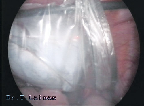
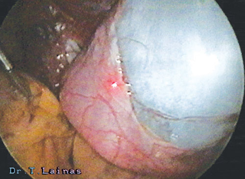
Ultrasound criteria correlate with the size, the location, the presence of ascites, the presences of cysts and solid areas in the same cyst, the sheer cyst or solid appearance, the thick walls, etc. Besides, the transvaginal Doppler ultrasound with colour flow imaging is a modern examination that offers significant assistance in determining blood flow in the cyst area. From the biochemical test, the C1-125 indicator is useful but has higher sensitivity and specificity for post-menopausal women.Laparoscopic criteria for benign findings (Mage G, 1987) correlate with the thickness of the wall, the vascularisation of the cyst, the colour of the fluid contents, the length of the same connection of the ovary and the appearance of the internal wall of the cyst. According to the pre-operative and peri-operative assessment of the cyst, laparoscopic surgery is performed. Special care is taken in the removal of the entire capsule (shell) of the cyst without it rupturing, as well as in the maintenance of the healthy ovarian tissue.In the case of rupturing, the cyst is thoroughly aspirated and its contents along with the fluids from washing out Pouch of Douglas are sent for cytological examination. A thorough examination of the internal of the cyst is then performed, in order to exclude any possible damage, the entire capsule of the cyst is removed and a quick biopsy is performed even when there is only suspicion of malignancy. For the removal of the cyst from the peritoneal cavity a special endoscopic pouch is used.
Types of ovarian cysts
The most common cysts in women of reproductive age are: the endometriotic (endometriomas), the dermoid and the cysts with clear content (bloody or mucoid).