Fertility


Διερεύνηση Γονιμότητας – Υπογονιμότητας

Human reproduction is the result of the union of a sperm with an egg. Eggs are produced in the ovaries and sperm in the testes. The union of the sperm with the egg will result in the embryo that will grow in the uterus so that the child is born.

The problems of infertility occur frequently and can appear at all ages.  Infertility is caused by female factors, male factors or mixed causes. A couple is described as “infertile” when systematically attempting to conceive for over a year without precaution or other methods of contraception, while being at a reproductive age.

The first question that should be answered at the beginning of infertility investigation and before treatment is whether despite adequate sexual intercourse, no pregnancy has occurred. The infertile couple should always be treated as a complete unit. Therefore, it is a prerequisite that both of the partners be present in their first visit to our Unit. A detailed record of your medical history and information from any previous attempts will be recorded in order to evaluate your specific conditions and to propose further diagnostic tests.

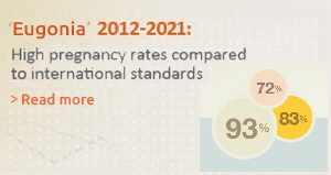
Recent scientific developments, combined with their individualized application (based on the principle of evidence based medicine) guarantee not only that the best treatment options are available to our patients, but also that their success rates will be high, reaching up to 88%.

Human reproduction is the result of the union of a spermatozoon with an egg. Eggs are produced in the ovaries and sperm in the testes. This union will result in an embryo that will develop in the uterus, resulting in the birth of a child.

If there is intercourse during the fertile days, the sperm will be deposited in the vaginal cavity. The sperm will then go through the cervical canal towards the uterine cavity and through the proximal tubal ostium to reach the fallopian tubes.

The fallopian tube will then receive the egg that was recently released with ovulation, via its fimbria. Sperm and egg meet in the ampulla of the fallopian tube and this is where the egg will be fertilised by only one sperm. The fertilised egg (zygote) will remain in the fallopian tube for 5-6 days, where it divides into 2, 4, 8 cells and then becomes a morula and a blastocyst.

As a blastocyst (an embryo with around 6-120 cells) it will then migrate towards the uterine cavity and implant by invading the endometrium where it continues to develop.


After conception

If there will be conception, the embryo begins to create its placenta at the point of implantation. The placenta secretes hormones, which stabilize the endometrium and prevent its degeneration. Menstruation does not come and the embryo continues its development normally, which the woman perceives with the delay of menstruation. Conversely, if one of the previous stages does not work (and at the right time), there is no pregnancy: the luteal phase of the cycle ends with endometrial cell degeneration (apoptosis) about 14 days after ovulation. The endometrium degenerates and falls with blood. The blood is drained through the cervix to the outside of the body, so the woman realize it as menstruation (period).


See also:

Normal fertilisation

Requirements of conception

Intrauterine insemination

What is IVF

The necessary requirements for conception to occur are the following:

  • The fallopian tubes must be unobstructed and functional to receive the egg from the ovary and allow it to meet with the sperm.
  • The sperm must have normal parameters of concentration, forward motility and viability to be able to reach the egg overcoming all the natural hurdles they meet in the way.
  • The passing of the sperm from the vagina to the fallopian tubes must be smooth with a friendly environment from the cervical mucus.
  • Ovulation must occur so that a mature egg is released from the follicle.
  • Sexual intercourse must take place during the fertile days.

It is known that:


  • Endometriosis significantly reduces fertility.
  • 25-30% of infertility cases are unexplained.


Before the onset of investigation and treatment of infertility we should answer an important question: has the frequency of sexual intercourse been adequate, despite which no pregnancy has occurred?

If the answer is yes and no conception has been achieved in a period of one year, then it is necessary to investigate the causes of subfertility in order to select the suitable treatment.

The philosophy of dealing with subfertility may include:

  • a "wait and see" period
  • undergoing tests to explore the causes of subfertility in order to select the suitable treatment
  • time course of treatment (intrauterine insemination, hysteroscopy-laparoscopy etc)
  • direct application of IVF

The diagnostic approach depends on whether the investigation involves a first visit to the doctor, a referral from a gynecologist, an andrologist, urologist, endocrinologist, possible previous infertility treatment (eg ovarian stimulation, insemination, fallopian tube surgery), and previous IVF.

The infertile couple should be investigated together as a unit. In the first visit it is necessary both of  them be present, according to international practice. Family and individual history, obstetric history (births, abortions, abortions, etc.), gynecological, surgical history (previous surgeries), age, weight, height, work, smoking, alcohol consumption, possible medication and previous tests (eg, semen analysis, hormone tests, ultrasound, hysterosalpingography, laparoscopy, hysteroscopy) are evaluated.


The diagnostic evaluation after 12-month infertility  includes an assessment of the following parameters:

  • Male Factor (sperm analysis: 2 or 3 different tests).
  • Check of ovulation and the luteal phase of the cycle (FSH, LH, PRL, E2 on days 2-6 of the cycle, progesterone on day 21 of the cycle, ovarian and endometrial ultrasound scanning).
  • Cervical factor: post-coital test.
  • Uterine factor (hysterosalpingography, hysteroscopy, ultrasound).
  • Salpingoperitoneal factor (hysterosalpingography and laparoscopy).
  • Endometriosis or other pathological condition of the genital system (laparoscopy).
  • Ovarian reserve (FSH, E2, ovarian volume, follicles in the 3rd day of the cycle).

Τhe definition of health remains unchanged since 1948. It is in the official constitutional text of the World Health Organization, which was accepted by the World Health Conference, signed on June 22, 1946 by representatives of 61 states in New York and entered into force on April 7, 1948. According to this definition, health is a state of complete physical, spiritual and social well-being, not simply the absence of illness or infirmity.

By this term, infertility is indeed a health disorder. The couple, who can not conceive  despite its desire, is not in "complete spiritual and social well-being," provided  that this is considered to be a social stigma and as a consequence this affects the spiritual and social well-being of infertile couples. Thus, infertile couples are "sick", but that does not mean that they need to be stigmatized, just as no one is stigmatized because of flu or myopia. Infertility does not have any particular symptoms, and "patients" do not seem to "suffer" from an obvious disease.

The problem of infertility occurs frequently and is always up-to-date.

Internationally, 15% of couples have difficulty in conceiving in general or in conceiving the desired number of children, according to World Health Organization (WHO). The probability of conception in a couple of reproductive age is 20% per month.

The rate of infertility remains stable over the centuries. In the 19th century, a study conducted in England, had showed that 1 out of 6 marriages weren’t reproductive (16%).

It is difficult to calculate accurately the percentage of infertile couples, because it requires a range of parameters and the exclusion of people who have been sterilized or use a contraceptive method but also those who do not wish to be conceived. In the US, there are approximately 10-15 million infertility couples, and 585,000 in Germany (1989 data). In Greece, there are approximately 300,000 couples.

This rate is stable in the last two decades, but the number of patients undergoing infertility treatment and the number of new fertility centers increase significantly.

Subfertility is the failure to conceive after 12 months of sexual intercourse without protection or use of contraceptives.

Infertility is the complete inability to conceive.

Monthly Fertility Rate (MFR) is the probability of conception in a menstrual cycle.

The human is not a particularly fertile mammal. It is estimated that in humans the MFR is 20%, while in baboons it is 80% and in rabbits 90%.

Despite the developments in fertility diagnosis, a percentage of 25-30% remains unexplained. In specific studies (Guzick et al.), routine tests in fertile couples (semen analysis, luteal phase determination, hysterosalpingography) allowed at least one infertility factor to be identified in 2/3 of the couples. In addition, infertile couples had normal test results in 5 of 32 cases, while only 10 out of 32 fertile couples had normal test results. Consequently, a pathological result does not necessarily imply infertility.

There is little data on the percentage of natural conception in patients with unexplained infertility who have exhausted conventional treatments. It is noteworthy that 5.9% of couples in the UK achieved that while they were in a waiting list for IVF treatment.

But what does unexplained infertility mean? If all the infertility factors have been tested and nothing abnormal has been found, why is there no conception?

Sadly, in many cases there is no certain answer to seemingly simple questions such as:

Did ovulation occur?

Was the oocyte mature and of good quality?

Did fertilization occurred?

Did the fertilized egg cleaved?

Did the embryo reached the blastocyst stage?

Was the endometrium capable of receive the embryo?

Was the embryo and endometrial interaction appropriate for successful implantation?

These are some questions that reflect the complexity of the mechanisms and procedures used during fertilization and embryo development. It is obvious that the factors studied in the investigation of infertility (eg hormonal profile, sperm quality, etc.) are very limited and impossible to identify all possible cause of infertility. For these reasons, unexplained infertility is a cause of infertility in 25-30% of couples.


Mild / moderate endometriosis and unexplained infertility

A review of 22 studies and 2026 patients, showed (Taylor and Collins) that mild and moderate forms of endometriosis affects fertility. Therefore, the role of minimal and moderate endometriosis in infertility is essentially unexplained, whereas severe endometriosis refers to tubal-peritoneal factors. Patients with endometriosis and unexplained infertility have generally the same prognosis in IVF cycles.


Age and unexplained infertility

The chance of diagnosing unexplained infertility increases significantly while woman’s age is increasing. Infertility is mainly associated with low ovarian reserve and oocyte quality. Especially for women over 40, the probability of natural conception is low, and extremely rarely over 44, even in women who have normal ovulation cycles.

Lifestyle may affect fertility. The high number of sex partners increases the risk of sexually transmitted diseases, resulting in a possible infection of the genital system. Also, smoking increases the risk of primary infertility. Increased consumption of alcohol is also related to the risk of female infertility. Body weight is associated with anovulation and disorders of the hypothalamic function. Excessive exercise and anxiety are also associated with infertility.

Furthermore, some data shows correlation of infertility with the environment at work. Despite the increased concern, men’s exposure to pesticides, lead and other heavy metals, show no effect on sperm parameters.

The cervical factor is almost exclusively associated with the quality of cervical mucus and its interaction with the sperm. Cervical mucus is secreted by glands found in and around cervix. Hormonal changes throughout a woman’s reproductive cycle change the amount and consistency of this mucus.

Its role is to nourish and help transport sperm through the cervix into the uterus. It is also a clue that ovulation is coming.

In some women, there are sperm antibodies in the mucus of the cervix. These create agglutinations and the sperm cannot reach the uterine cavity, even in the ovulation phase (fertile days). The hostility of cervical mucus is the main cause of infertility in 9-15% of couples.

The main way of testing the mucus is the postcoital test (PCT), but this test is very controversial. Its effectiveness is disappointing in clinical practice and its predictive value is considered by many to be limited. Typically, positive PCT indicates the presence of a sufficient number of mobile sperm in mucus 4-10 hours after contact, and negative PCT shows the absence of sperm. Practically, if more than 50 sperm with progressive motility per field of vision is found in PCT, in the mucus, 9-24 hours after contact, male factor can be excluded as cause of infertility.

A couple’s infertility can be attributed to either the woman, the man or both. Usual causes of infertility include:

  • Issues with the male partner’s sperm
  • Ovulation disorders
  • Cervical disorders, such as “hostile” cervical mucus
  • Uterine disorders
  • Tubal disorders (blockage or adhesions)
  • Endometriosis
  • Age
  • Life style factors (smoking, alcohol, work environment)
  • Unknown causes (which may represent as much as 25-30% of cases).

Retrograde ejaculation is a rare disorder in which sperm is abnormally ejaculated into the bladder and therefore is found in the man's urine.

This problem can be solved via IVF, with the isolation of sperm from the man's urine. However, the number and quality of sperm cells is usually low and therefore ICSI is the preferred method of fertilization.  Specialized embryologists are essential for success.

A general semen analysis includes:

  • Macroscopic evaluation of the sample, such as volume, liquefaction time, pH.
  • Microscopic evaluation, consisting of:
  1. a) Total sperm count (concentration), which is expressed as millions of spermatozoa / ml
  2. b) Sperm motility, which is split into 4 categories (rapid progressive motility, slow progressive motility, non-progressive motility and immotile).
  3. c) Sperm morphology (head, midpiece and tail)
  4. d) Count of white blood cells.


According to the criteria of the World Health Organization (WHO), a sperm sample with the following characteristics is considered normal.

  • Volume: at least 1,5 ml
  • Sperm count: at least 000.000 /ml
  • Progressive motility: At least 32%, within 1 hour.
  • Morphology: At least 4% of spermatozoa should be normal.


Sperm disorders

Oligospermia: Low number of sperm cells (less than 15 million/ml or 39 million total).

Asthenospermia: Low progressive motility (less than 32%).

Teratospermai: Low percentage of morphologically normal sperm cell (less than 4%).

Oligoasthenoteratospermia (ΟΑT): Combination of low numbers, low motility and poor morphology. This is one of the most common disorders.

Azoospermia: Complete absence of sperm cell in the ejaculate.

Aspermia: Lack of ejaculation.

Azoospermia is the complete absence of sperm cell in the ejaculate.

Obstructive azoospermia (48% of cases) is caused by a blockage of both posttesticular genital tracts.

Non obstructive azoospermia is indicative of a lack of production of sperm cell by the testes and can be caused by chromosomal abnormalities, infections, endocrine or metabolic disorders or environmental causes.

It usually involves a non-regular menstrual cycle, combined with high levels of gonadotrophins and normal prolactin levels. These signs usually point towards ovarian failure combined with elevated FSH (> 20 mIU/ml) and low oestradiol. When this is occurs in women under 30 years of age it is called premature ovarian failure

  • Premature ovarian failure occurs in 1% of all cases of infertility an in 10% of women with amenorrhea.
  • Poor responders are women whose follicles do not develop despite being treated with gonadotrophins.Around 2/5 women with premature ovarian failure are also poor responders.
  • Primary gonadal dysgenesis is a rare condition in which follicles are not formed during the childs embryonic development. The absence of follicles can only be treated via egg donation.
  • Empty follicle syndrome is a rare condition, which is diagnosed via IVF when two or more cycles fail to gain any oocytes from the egg collection despite the presence of seemingly mature follicles.

The hypothalamus and pituitary gland are the main centers controlling the hormones GnRH, FSH and LH, which in turn affect the ovary in order to begin and maintain a woman’s cycle. If one or more of these hormones is not being produced adequately then ovulation disorders are unavoidable, leading to infertility

The main causes of hypogonadotropic-hypogonadism are:

  • Kallman Syndrome: A rare condition which causes the lack of GnRH production, leading to anovulation. It is genetically transmited.
  • Severe weight loss (Anorexia nervosa): This is a psychosomatic disorder that is often found in teenagers and more commonly in girls rather than boys. It results in large weight loss and this can disturb the production of GnRH leading to amenorrhea.
  • Obesity: Obesity is associated with low SHBG levels, high oestrogen and androgen levels which disturb the function of FSH and LH leading to hypogonadism.
  • Intense physical exercise: Women who exercise excessively have a higher chance of anovulation due to lower levels of FSH and LH and higher levels of prolactin. A study found that two thirds of female runners have small ovulation phases in their cycles or complete anovulation.

In most cases when normal body weight has returned, fertility is restored without further treatment needed. Therefore it is important for women with high or low body weight to follow a correct diet before commencing treatment.

  • Hypothalamus injury: The hypothalamus can be damaged by some tumors, tuberculosis, head injuries, radio-treatment etc. This causes disturbances to the production of GnRH, which leads to infertility.

Sheehan Syndrome: A rare condition which is characterized by a partial or total pituitary necrosis caused by bleeding and clotting of blood vessels. It can occur during labor and can be diagnosed by a difficulty to produce milk, hypotension and amenorrhea.

Hyperprolactinaemia is diagnosed when prolactin blood levels are higher than 20 ng/ml. These high levels inhibit the production on GnRH which results in the lowering of FSH and LH. The most common causes of hyperprolactinaemia are: stress , PCOS, hyperthyroidism, some types of medication (such as antihistamines, a few contraceptive pills, oestrogen, dopamine antagonists and amphetamines), breast operations and herpes infections.

The main symptoms are ovulation disorders leading to infertility and low libido. Hyperprolactinaemia can be accompanied by prolactinomata (tumors in or near the pituitary) and, rarely, Cushing syndrome may also be present. In a small number of women, hyperprolactinaemia has been associated with hypothyroidism due to low levels of thyroxin causing high levels of TSH and prolactin. The most effective treatments are dopamine agonists and hypothyroidism medication.

Polycystic ovary syndrome (PCOS) is the most common endorine condition in women of reproductive age and also the most common ovulation disorder. Dispite huge progress in detecting and treating PCOS the cause of the condition is complicated and not well understood.

Young women with PCOS can exhibit metabolic disorders, such as insulin resistance, which have been linked to type 2 diabetes later on in life. The main problem caused by PCOS is ovulation anomalies and therefore infertility. Even so, many women with PCOS have normal cycles and go on to conceive naturally. One study, however, found that 75% of women with PCOS had ovulation disorders and infertility, while abnormal cycle lengths and/or anovualtion was present in 80% of PCOS women (Van Santbrink et al., 1997).

Treatment

In 2007 in Thessaloniki, an international consensus was reached regarding treatment strategies for PCOS. A first option for treatment is the use of clomifene citrate to induce ovulation. A second choice is gonadotropin administration if the first option fails. Gonadotropin administration must be monitored closely, due to the risk of ovarian hyperstimulation (OHSS). Metformin administration can lower insulin levels, but this method is no longer recommended. Finally, weight loss and physical exercise are highly recommended, as  they increase the effectiveness of treatment and alleviate some of the symptoms, such as type 2 diabetes.

IVF is the most effective method of dealing with the infertility associated with PCOS. Use of GnRH analogues (agonists and antagonists), combined with gonadotropins in order to undergo IVF can lead to very high pregnancy rates, while close monitoring is required to minimise the risk of OHSS. Women with PCOS undergoing IVF are much more likely to develop OHSS than women without PCOS. OHSS can become a serious health concern if not dealt with properly.  At Eugonia we are world leaders in OHSS prevention and treatment and have contributed significantly to modern international strategies of OHSS management. We have published the largest study regarding women with PCOS that have undergone IVF and found that the use of a flexible GnRH antagonist protocol is related to significantly lower incidence of OHSS compared to the older long protocol, while maintaining high pregnancy rates. Therefore an antagonist protocol can be considered as the best option for women with PCOS undergoing IVF.

When the tube is blocked at it's end (infundibulum), it can trap fluid, and is called a hydrosalpinx, which can cause a chronic inflammation. The tube is non functional and, in additon, toxic substances created by the inflammation may obstruct the implantation of the embryo when it is transfered into the uterus during IVF procedures.

Therefore, surgical treatment of hydrosalpinx is important and involves either opening the blocked end where possible or removing the fallopian tube in more serious cases.

Chlamydia is an asymptomatic sexually transmitted disease. Due to a lack of serious symptoms, most women are unaware of the inflammation until they are faced with infertility.

If the inflammation spreads to the fallopian this may induce a fever combine with  pelvic pain. Eventually, adhesions caused by the inflammation will cause tubal blockage and infertility.

Mild damage to the fallopian tubes can affect the collection of the egg or its fertilisation and transport. Adhesions can disturb the anatomical relationship of the fallopian tube with the ovary.

Serious damage to the fallopian tubes usually leads to tubal blockage and infertility. Causes can include:

  • Tubal inflammations,
  • Other inflammations of the area such as appendicitis
  • Inflammatory complications due to surgical operations in the area.
  • The most common cause of blockage is chlamydia or gonorrhea infection.

Proposed mechanisms through which endometriosis may affects women's fertility are the following:

  • Mechanically, via adhesions, immotile ovaries, tubal blockage etc.
  • Ovulation disorders such as anovulation, poor luteal phase and/or luteinized unruptured follicle syndrome.
  • Dysfunctional fallopian tubes due to toxic factor caused by the condition, such as prostagladins.
  • Reduced implantation potential of the embryo due to integrins and other embryotoxic factors produced by the condition.
  • Modified immune response due to high T, B and NK counts.
  • Pelvic inflammation which can lead to high levels of IL1, TNF and prostaglandins.

Women with poor response are not necessarily infertile, if another infertility factor does not coexist.

The ovaries of these women have a markedly reduced response to gonadotrophins which are administered to induce multiple follicular development.

See more

It is well known that the maximum fertility rate in women is at the age of 24 (fertility rate 86%), while the percentage in women 25-30 years old is about 78%. At age 30-34, fertility begins to decline (63%) and after the age of 35, it is more noticeable (52%). Natural conception is rare after the age of 45 (less than 0.1%).

Accurate estimation of ovarian reserve helps to predict responsiveness to stimulation and to properly predict the likelihood of pregnancy success in IVF cycles. The tests for the determination of ovarian reserve are FSH, E2, AMH and vaginal ultrasound.

Endometrial scars and atrophic, fine endometrium are usually the result of trauma and are associated with decreased blastocyst implantation.

The pathology of the isthmus and endocervical cavity can be linked to infertility. The obstruction creates a mechanical infertility factor, while scars, constrictions, encapsulated cysts (usually result of traumatic procedures) are associated with the production and consistency of cervical mucus.

It is a rare condition associated with secondary infertility. Less than two hundred cases have been reported in the international literature.

See more

Endometritis is an inflammation of the endometrium and may be associated with unexplained infertility (14.3%) or repeated miscarriages (22.6%), (Hamou, 1991).

Fibroids (or leiomyomas) are benign tumors of the uterus and are classified as submucosal, intramural and subserosal. Intramural and subserosal fibroids are usually asymptomatic, unless they are very large in size causing effects of pressure.

See more

Endometrial polyps are usually asyptomatic, benign tumors of the uterus and may be responsible for the anomalous uterine haemorrhage and can be found during intravaginal ultrasound or hysterosalpingography.

See more

Endometrial adhesions may be a cause of infertility (tubal - peritoneal cause) and probably of pelvic pain.

See more

Congenital anomalies of the uterus (Mullerian duct anomalies) do not seem to directly correlate with subfertility issues, except for uterus aplasia and its variations, as well as cervical and vaginal atresia and the complete transverse vaginal septums.

See more

During your first appointment, you will be welcomed by the coordinator of the unit. The consultation will be conducted in three phases:

A midwife will initially take a detailed record of your medical history.
You will then meet the Medical Director of the Unit, with whom you will have the opportunity to discuss the recommended diagnostic and therapeutic strategy. The doctor will note in your history all necessary tests required and the treatment protocol recommended for you.
Finally, the midwife will explain and provide you with details about when and how the required tests will be conducted. You will also be given prescriptions for any medication necessary, instructions about the treatment protocol, dosage, time and explanation how to take the medication, and also consent forms and instructions about your next appointment with us.

Please note that this first appointment may be long, depending on the information that must be recorded and any queries you might have about the treatment that you wish to discuss with the doctor (usually 1 - 2 hours).

Endometrial biopsy

In the past, this was a routine, but nowadays its usefulness has been questioned.

Samples can be collected by different ways and on different days of the cycle, depending on the findings (eg following hysteroscopy) and the phase of the cycle that must be checked. The aim is to histologically determine the phase of the cycle, especially the luteal phase, when anomalies in its progression are suspected (e.g. luteal phase deficiency).

In rare cases, the biopsy shortly after ovulation allows the examination of specific formations of the endometrial cells (pinopodes) using  electron microscopy, that are considered to be related to embryo implantation. This test is still considered experimental and is generally not recommended in everyday clinical practice.


Thermometric diagram

It was widely applied in the past to predict ovulation. It has now been replaced with ultrasound and hormonal testing


Cervical mucus assessment

It is an old diagnostic method of ovulation.


Salpingoscopy

It is an endoscopic method of assessing the epithelium and tubal lumen. The development of IVF overturned the possibility of its development and wider application.

Cystic fibrosis is a metabolic disorder characterized by generalized dysfunction of the glands and overproduction of mucus with high viscosity. This leads to chronic bronchial obstruction and respiratory tract infections, as well as disorders of pancreatic excretory function. In men with cystic fibrosis, infertility is very common.

In Europe its frequency is one in 2000 births and is considered the most common congenital metabolic disorder. Genetically, it is an autosomal recessive hereditary disorder. More than 600 mutations in cystic fibrosis are known, the most common being ΔF-508.

It is understandable that testing should be done in couples with relatives who suffer from cystic fibrosis. It is also advisable to test for cystic fibrosis couples with severe male infertility. If one of the two candidate parents bears the disease, preimplantation genetic diagnosis (PGD) may be employed to diagnose the embryo(s) prior to the embryo transfer.

The depiction of an individual’s entire chromosomal complement is called a karyotype and a normal karyotype consists of 46 chromosomes (23pairs).

Karyotype testing can help detect numerical and structural chromosomal abnormalities.

It is recommended in cases of repeated miscarriages and abortions (> 3), oligoasthenospermia, in repeated IVF failed attempts and if a fetus with a chromosomal anomaly is delivered.

Thrombophilia is an abnormality of blood coagulation that increases the risk of thrombosis (blood clots within blood vessels). It can be congenital or acquired.  According to statistics, 10-20% of the population suffers from thrombophilia and may develop thromboembolic events such as stroke, myocardial infarction in deep vein thrombosis, and pulmonary embolism.

Often these thrombophilic conditions are responsible for pregnancy complications such as: first trimester miscarriages, pre-eclampsia, delayed fetal development, placental abruption and fetal death.

Common types of hereditary thrombophilia are: deficiency in proteins C and S, antithrombin deficiency, factor V Leiden mutation, prothrombin gene (G20210A) mutation and MTHFR gene mutation.

Hormonal testing is an irreplaceable tool in investigating and treating infertility.

Ovulation problems are a major cause of female infertility. Follicular development, selection and follicle maturation, as well as maturation and oocyte ovulation, are all processes regulated by complex hormonal mechanisms. Measurements of hormone levels at certain days of the cycle can help diagnose abnormalities in the cycle, ovulation, etc.

Furthermore, the measurement of FSH and E2 levels at the beginning of the cycle allows us to roughly determine the so-called "biological age" of the ovary and its ability to develop follicles.

In special cases, hormone testing is supplemented with measurements of testosterone, free testosterone, Δ4- Androstenedione, dehydroepiandrosterone (DHEA), DHEA sulfate, sex hormone-binding globulin (SHGB) and 17-hydroxyprogesterone (17OH-PRG). In thyroid disorders, an endocrinologist’s examination is considered necessary and, depending on his/her recommendations, TSH, T3, T4, anti-thyroid antibodies, thyroid ultrasound, etc. are performed.

Hysterosalpingography, also known as uterosalpingography is a series of static radiographs obtained after injecting a radio-opaque fluid into the cervical canal. The fluid fills the lumen of the cervix, the uterine cavity and finally flows through the fallopian tubes into the peritoneum.

Hysterosalpingography is performed on the 10th-11th day of the cycle and is mainly used to assess the cervical lumen, the shape of the uterus and the shape and patency of the fallopian tubes.  It is also used to investigate congenital anomalies of the uterus, cervix and fallopian tubes and determine the presence and severity of tumor masses, adhesions and uterine fibroids. Hysterosalpingography can also diagnose and occasionally open blocked fallopian tubes.

The ultrasound examination that may be performed vaginally or abdominally is a safe and painless method of examination. The contribution of the ultrasound to the diagnosis of normal and pathological conditions of the genital system is invaluable. We can estimate:

  • The anatomical position, size and texture of the uterus and ovaries
  • The thickness and texture of the endometrium
  • The biological age of the ovaries (ovarian reserve)
  • The presence and size of the follicles or the corpus luteum in the ovary
  • Ovulation
  • The presence of cysts, tumors or lesions in the ovary or uterus
  • The onset of pregnancy. In particular, the number and size of the gestational sac(s), the presence of the fetus, his cardiac functions, etc.
  • Early diagnosis of ectopic pregnancy

Finally, the ultrasound is a very valuable diagnostic tool during pregnancy (nuchal translucency ultrasound, estimation of fetal biophysical parameters, etc.).

The ovary is a "pool" of follicles at various developmental stages. The term “ovarian reserve” refers to the number of follicles found in the ovaries and represents the ovaries’ biological age.

See more

By monitoring follicular growth every two days, it is possible to predict the rupture of the follicle (ovulation), which is confirmed by a final ultrasound. The thickness of the endometrium is also recorded. Ultrasound examination combined with blood hormonal levels help determine the possible day of ovulation, hence the woman’s "fertile day".

Semen analysis is sometimes not enough to assess sperm ability to fertilise. Therefore, there are many other specific sperm tests to detect sperm function. Such a test is the Hyaluronan Binding Assay (HBA) which assesses sperm maturity and fertility. It is based on the principle that hyaluronic acid selectively binds to mature spermatozoa that carry an intact acrosome and normal morphology. Hyaluronic acid is normally found in the extracellular matrix of the granulosa cells which surround the ovum. It forms a layer which creates a natural obstacle, as it allows only mature spermatozoa, which have special receptors, to bind and cause lysis of hyaluronic acid. In this way they manage to reach and penetrate zona pellucida, in order to achieve the acrosome reaction and consequently the egg fertilization.

A low-binding HBA analysis corresponds to a low number of mature spermatozoa and is therefore a prognosis of infertility. Conversely, hyaluronic acid-binding spermatozoa are able to interact with the egg and  they have high integrity of their genome.

Thus, samples with > = 80% binding are considered to have normal maturity and function, whereas binding <80% indicates reduced maturity and function.

In case of an abnormal HBA test, fertilization can be done using the new technique PICSI.

Sperm DNA fragmentation

The integrity of genetic material in the sperm is crucial for successful fertilisation and normal embryo development.  Male subfertility, IVF failure and miscarriage may be due to sperm DNA fragmentation. The major contributing factors for DNA fragmentation are the environmental toxins and the reactive oxygen species(ROS).

Recent studies have shown that a semen analysis is not indicative of the presence of high percentage of apoptotic sperm DNA, and several cases of unexplained male infertility have been justified following the examination of DNA fragmentation.


Cytogenetic sperm examination

International literature reports that infertile men with normal peripheral blood karyotype have a higher incidence of chromosomal abnormalities in spermatozoa compared to men with normal sperm parameters. Indicatively, the most common chromosomal abnormalities are the abnormal number of sex chromosomes or structural chromosomal abnormalities. As a result, an increased number of embryos with chromosomal abnormalities can be created, reducing the chances of IVF success.


The criteria for sperm cytogenetic examination and DNA fragmentation include:

  • Recurrent biochemical / retrograde pregnancies in women under 35 years old with normal karyotype
  • Bad quality embryos in women under 35 with normal  karyotype
  • Inability of the sperm to fertilize
  • Men undergoing radiotherapy or chemotherapy
  • Men with oligoasthenoteratozoospermia
  • Unexplained infertility

The investigation of severe oligoasthenoteratotterospermia (OAT) and azoospermia includes:

  • Hormone test(FSH, LH, PRL, Testo, TSH),
  • Testicular ultrasound with Doppler,
  • Karyotype
  • Test for micro- deletions in chromosome Y,
  • Cystic fibrosis test
  • Testicular biopsy and histological examination.

In special cases, collaboration with specialized urologists or endocrinologists in the diagnosis and treatment of male infertility is beneficial.

Sperm cryopreservation is an important component of fertility management, and much of its successful application seems to affect the reproductive outcome of ART.

Sperm should be cryopreserved:

  • When there is a risk of reproductive ability loss (testis removal, chemotherapy, radiotherapy.)
  • When the male partner will be absent on the day of oocyte retrieval.
  • In cases of surgical sperm recovery.
  • In cases of sperm recovery using electroejaculation.
  • In cases of ejaculation problems.
  • In cases of progressively declining sperm quality.
  • Before sterilization by vasectomy.

Sperm culture – Antibiotic sensitivity

Sperm culture allows the detection of a specific pathogen microorganism. Then, the  identification of these bacteria and the study of their sensitivity to various antibiotics take place. In case that the culture is positive, medical treatment must be administrated and the examination must be repeated in order to confirm that there are no bacteria after the treatment. Abstinence 2-5 days is recommended before the examination.

The separation of spermatozoa from seminal plasma is needed for a variety of purposes, such as diagnostic tests of function and therapeutic recovery for insemination and assisted reproductive technologies. Density-gradient centrifugation and swim-up are the preferable methods for sperm preparation.


Density-gradient centrifugation

During normal conception, spermatozoa are separated from the seminal plasma and acquire specific characteristics when passing through the uterine cervix. In the laboratory, a special method allows us to mimic this stage.

The method uses centrifugation of seminal plasma over density-gradients consisting of colloidal silica coated with silane, which separates cells by their density.

Seminal plasma, white blood cells, debris, as well as bacteria and fungus are retained in the colloidal intermediate layers along with immotile spermatozoa. Motile spermatozoa swim actively through the gradient material to form a soft pellet at the bottom of the tube. After discarding the supernatants,  the pellet is washed to remove the colloid. The procedure lasts about 60 minutes and is used for diagnostic purposes or before intrauterine insemination or IVF/ICSI.

The sperm concentration and motility are assessed before and after preparation. As a rule, the number decreases, but the motility and morphology  are improved by this technique. The results of this test determine the IVF treatment (insemination, IVF, ICSI).

In very special cases, spermatozoa do not withstand this treatment, which is extremely selective. Then other methods are used, such as the swim-up method. This method is less selective and secure and, therefore, should be applied when there is no other option.


Swim-up

The sperm sample is diluted with culture medium and centrifuged. The supernatant is discarded, the pellet with the spermatozoa and other cells are resuspended to a small volume of culture medium, supplemented with about 1 ml of additional medium and placed in an incubator. The spermatozoa "swim" within the supernatant. Only motile and rapid motile spermatozoa are contained in the uppermost medium, which is carefully removed. The method is currently rarely used and it is not recommended if the motility is low and the sample contains many leukocytes.

The presence of sperm  antibodies is generally a recognized cause of infertility, although their mode of action is not well known. These antibodies cause sperm agglutination that influences  their motility, their interaction with cervical mucus, and penetration in the oocyte, resulting in fertilization failure. Also, it has been shown that sperm antibodies affect the embryos development, particularly in the 6-8 cell stage on the 3rd day of culture or before the blastocyst’s formation on the 5th or 6th day. These antibodies can be produced after inflammation, trauma or surgery.

The most common tests are: the MAR test (Mixed Antiglobulin Reaction, the SPAT test (Sperm Agglutination Test) and the immunobead test.

According to World Health Organization (WHO) guidelines, the presence of sperm antibodies may have negative effects on pregnancy when 50% or more of the sample's motile spermatozoa are agglutinated. In cases in which the test indicates the presence of sperm antibodies> 50%, ICSI is recommended.

Eugonia - Assisted Reproduction Unit
Konstantinou Ventiri 7(HILTON), 11528 Athens

  • Email: info@eugonia.com.gr
  • Τel.: +30 210 723 6333
  • Fax: +30 210 721 3623

Member of:

Google Reviews

Photo gallery

© Copyright 2026 «Ευγονία» - Μονάδα Εξωσωματικής Γονιμοποίησης στην Αθήνα Ανάπτυξη εφαρμογής NetValue. Με την επιφύλαξη παντός δικαιώματος.
Menu